
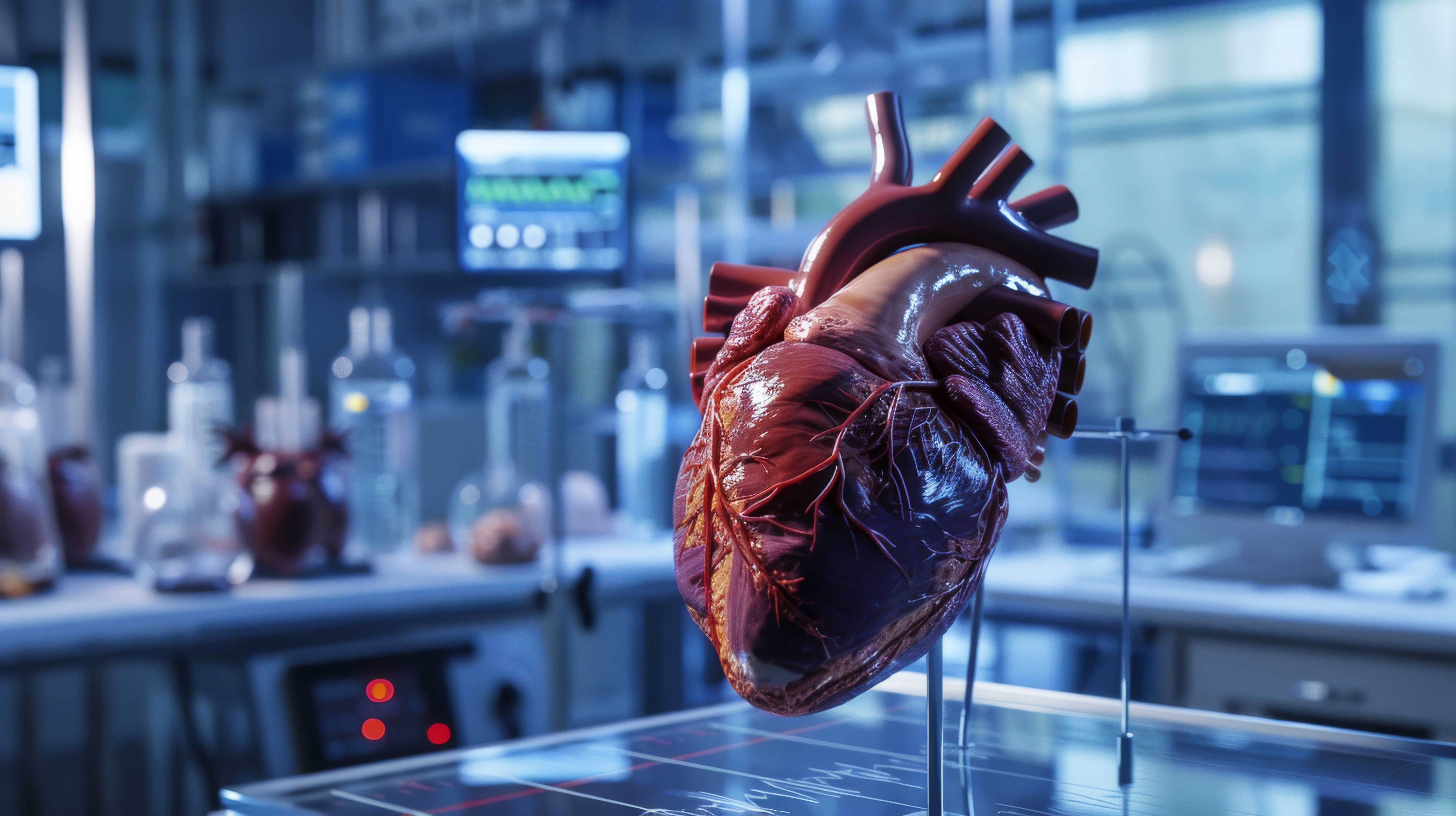
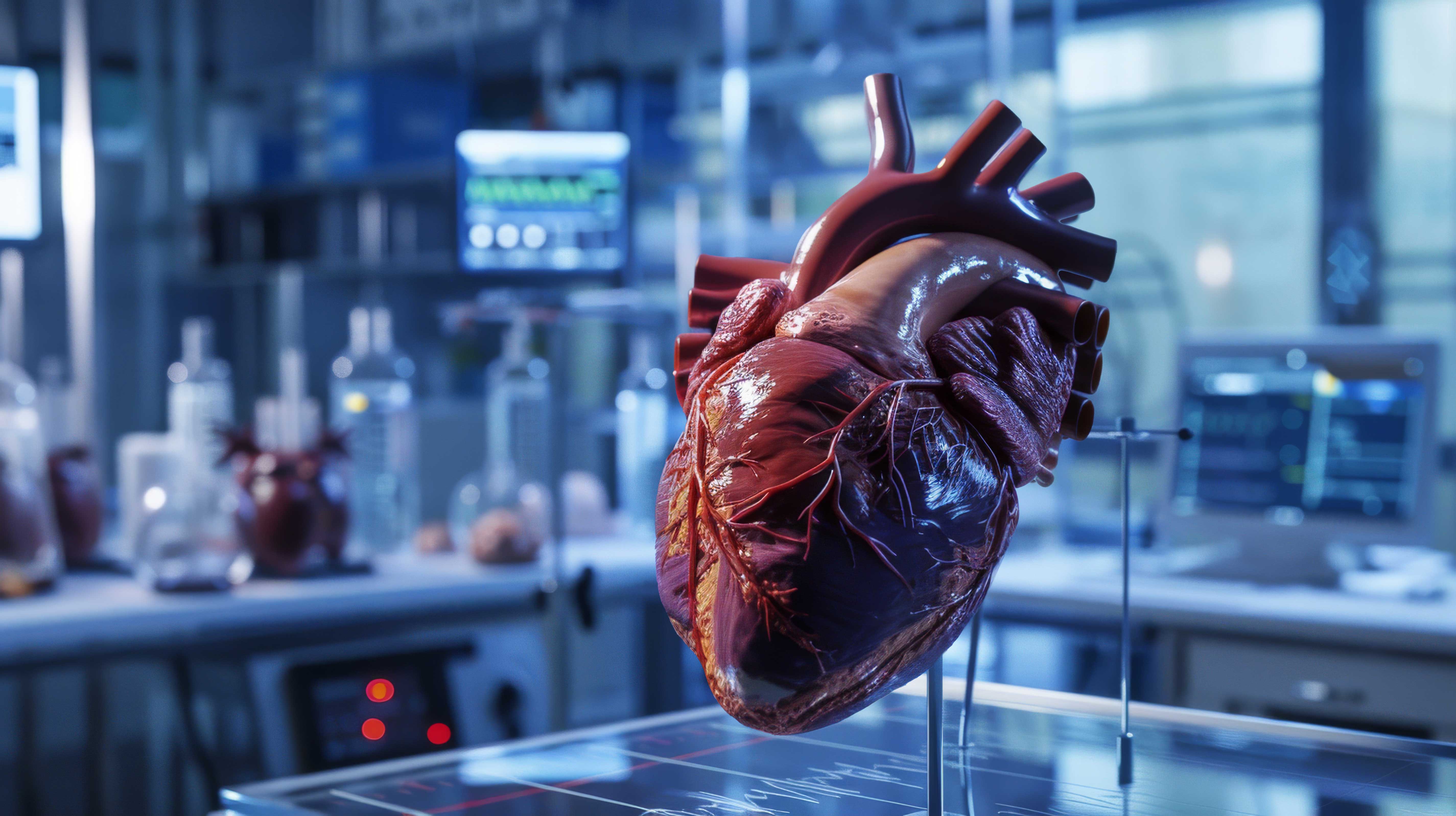
The heart after a heart attack
The story of a battle waged in the mitochondria
Wroclaw Medical University


Restoring blood flow in a blocked coronary artery after a heart attack now allows cardiologists to save a portion of the heart muscle—something that seemed impossible just a short time ago. The paradox, however, is that the very moment blood returns to oxygen-deprived tissue can trigger another wave of damage. This process, called ischemic-reperfusion injury, has remained one of the greatest challenges in modern cardiology for years.
Research by a team of scientists from Wroclaw Medical University, in collaboration with researchers from the Lower Silesian Center for Oncology, Pulmonology, and Hematology, and the University of Saskatchewan in Canada, points to one molecular mechanism underlying this phenomenon. The authors analyzed the role of a specific MMP-2 isoform in heart damage following ischemia. The study’s findings suggest that there may be a specific “biological switch” in the heart that determines whether a cell survives the moment blood flow returns—or begins a process leading to its own death.
A chemical storm
During a heart attack, a portion of the heart muscle is cut off from its oxygen supply. The cells then switch to emergency mode. Their metabolism slows down, energy production drops, and the mitochondria—the tiny cellular “power plants”—begin to function erratically.
When blood flow is restored, for example, during angioplasty, oxygen returns to the cells. This might seem like good news. In reality, however, a large amount of reactive oxygen species—very aggressive chemical molecules—is produced in cells. This phenomenon is called oxidative stress. This process can be compared to a situation in the power grid—after a long power outage, the sudden restoration of voltage causes power surges that damage part of the system. In heart cells, reactive oxygen species play a similar role.
As explained by Prof. Iwona Bil-Lula, the study’s author
- In our study using an ex vivo model of a rat heart subjected to ischemia and reperfusion, we demonstrated that activation of the mitochondrial isoform NTT-MMP-2 under these conditions leads to increased oxidative stress, mitochondrial dysfunction, and the activation of apoptotic pathways in cardiomyocytes, resulting in damage and impaired mechanical function of the heart.

Prof. dr hab. Iwona Dorota Bil-Lula Department of Laboratory Diagnostics Division of Clinical Chemistry and Laboratory Hematology Wroclaw Medical University
Prof. dr hab. Iwona Dorota Bil-Lula Department of Laboratory Diagnostics Division of Clinical Chemistry and Laboratory Hematology Wroclaw Medical University


The variable nature of the MMP-2 enzyme
One protein that responds to oxidative stress is the matrix metalloproteinase MMP-2. Under normal conditions, this enzyme participates in tissue remodeling. Among other things, it helps break down components of the extracellular matrix. However, under conditions of oxidative stress, an atypical, truncated version of it appears: NTT-MMP-2. Unlike the classic MMP-2 form, it remains inside the cell, particularly near the mitochondria. That is where the problem begins.
Researchers have shown that during ischemia and reperfusion, the activity of this mitochondrial enzyme increases dramatically. At the same time, reactive oxygen species production increases, inflammatory signals emerge, and cells begin to activate the cell death program.
In other words, NTT-MMP-2 may act as an internal catalyst for a destructive chemical reaction. As Prof. Bil-Lula emphasizes, MMP-2 is continuously produced by cells, but its levels increase under stress. It then begins to break down sarcomere proteins, which impairs muscle contractility.


Podcast: The heart after a heart attack
The heart in the laboratory
To better understand this process, the researchers used an experimental model frequently employed in cardiological research: the so-called Langendorff perfusion of an isolated rat heart. This method, developed in the late 19th century by the German physiologist Oscar Langendorff, remains one of the most important tools for studying heart function in laboratory conditions. In this setup, the heart is removed from the body but continues to beat—kept alive by the artificial circulation of a special nutrient solution that supplies the cells with oxygen and energy. This solution flows through the coronary vessels in a controlled manner, mimicking natural blood circulation.
The greatest advantage of this model is the ability to precisely control experimental conditions. Researchers can adjust the oxygen concentration, temperature, perfusion pressure, or chemical composition of the fluid, and can also introduce specific substances at precisely planned times. This allows them to observe the heart’s response to specific stimuli almost in real time—without interference from other organs or body systems.
The study simulated the three typical stages of a heart attack. First, the heart functioned under stable conditions, with a constant supply of oxygen and nutrients. Next, the perfusion fluid supply was stopped for a period of time, simulating ischemia—a situation analogous to the occlusion of a coronary artery in a patient with a heart attack. After this period, circulation was restored, initiating the reperfusion phase, i.e., the return of oxygen to previously oxygen-deprived tissue.
It was precisely this moment that proved to be the most dramatic from the perspective of cell biology. During reperfusion, the researchers observed a sharp increase in mitochondrial NTT-MMP-2 activity in cardiac muscle cells. At the same time, levels of markers indicating cell damage rose, and the heart’s ability to contract effectively declined significantly. Analysis of the data revealed a clear correlation: the higher the mitochondrial enzyme activity, the worse the heart’s recovery of normal rhythm and contractile force.
In practice, this meant that the moment the heart was once again receiving oxygen and nutrients—a moment that, in clinical settings, signifies the patient’s survival—a series of biochemical reactions promoting cell damage was simultaneously triggered within the cells.


An attempt to silence the enzyme
The most interesting part of the experiment involved an attempt to stop this destructive chain reaction before it could cause serious damage to the cells. The researchers turned to a tool that has become increasingly common in molecular biology and experimental medicine in recent years: siRNA (small interfering RNA) technology. These are very short fragments of genetic material designed to silence specific genes. In practice, they act as a precise “switch.” Once introduced into a cell, they recognize a specific RNA molecule responsible for producing a given protein and prevent its translation.
In this study, the siRNA was designed to reduce MMP-2 production, thereby lowering the amount of its truncated mitochondrial form—NTT-MMP-2. The aim was to determine whether inhibiting this single piece of the molecular puzzle could influence the entire process of heart damage during reperfusion.
The effect was not only noticeable but also statistically significant.
After partially inhibiting the enzyme’s activity, NTT-MMP-2 activity in heart cell mitochondria decreased, the production of reactive oxygen species—one of the main sources of oxidative stress—was significantly reduced, and the heart regained a better ability to contract and generate pressure. In other words, inhibiting the activity of a single enzyme led to an improvement in the mechanical function of the entire organ.
At the same time, the researchers observed a decrease in the levels of molecules associated with inflammatory processes and signals leading to apoptosis, or programmed cell death. These included, among others, components of signaling pathways activated in mitochondria under conditions of severe cellular stress. It is precisely these signals that constitute a biological tipping point—the moment when a cell ceases to attempt adaptation and begins to activate mechanisms leading to its own elimination.
As the study’s author explains:
“The results presented in the study suggest that inhibiting the activity of MMP-2/9, including the NTT-MMP-2 isoform, may represent a promising therapeutic approach for limiting myocardial damage associated with ischemia and reperfusion,” notes Prof. Bil-Lula.
Although the experiment was conducted in a laboratory model, the results reveal something significant. The processes leading to heart damage after a heart attack are not inevitable. To some extent, they can be modulated at the molecular level by interfering with specific enzymes and signaling pathways within cells.
The interleukin paradox
However, the experiment also yielded a surprising observation. After inhibiting MMP-2, levels of interleukin-6 increased—a signaling molecule belonging to the cytokine family, which plays a key role in communication between immune system cells. At first glance, this might seem like an undesirable effect. Interleukin-6 is often associated with inflammatory processes, and in many diseases—from infections to autoimmune disorders—elevated levels of it are a sign of an activated inflammatory response.
But in biology, things are rarely that simple. A growing body of research shows that IL-6 is a molecule with a dual nature. Depending on the biological context, the timing of its action, and the type of cells involved, it can both exacerbate inflammation and trigger protective mechanisms. In the case of myocardial ischemia, its role is particularly complex.
As Prof. Bil-Lula explains:
- Interleukin-6 is a cytokine with complex effects—in addition to its pro-inflammatory functions, it can also activate protective signaling pathways, including those related to cell survival and adaptation to oxidative stress.
In the experiment under analysis, the increase in IL-6 levels occurred precisely when MMP-2 inhibition began to limit damage to heart cells. Furthermore, increased expression of this cytokine was associated with less myocardial damage and improved cardiac mechanical function after reperfusion. This suggests that heart cells, which were partially protected from NTT-MMP-2, may have simultaneously activated additional adaptive mechanisms.
- Thus, the increase in IL-6 expression following siRNA administration may reflect a compensatory mechanism aimed at cardioprotection, the researcher concludes.
From a biological standpoint, such a mechanism is not surprising. Interleukin-6 can activate multiple signaling pathways that support cell survival under severe stress. In the short term following an ischemic episode, it can, among other things, stabilize mitochondrial function, limit apoptotic processes, and enhance cellular adaptation to oxidative stress. As a result, cells have a better chance of surviving the sudden return of oxygen, which normally triggers a cascade of damage.
In the study, the increase in IL-6 was thus associated with less heart damage and better myocardial mechanical function. It is therefore possible that the observed effect is part of a natural, compensatory cellular response—a kind of biological “rescue system” that activates when cells attempt to survive extreme stress.

A new therapeutic target
The authors emphasize that their experiment also has limitations. The isolated heart model does not fully reflect the complex processes occurring in the human body, and the number of samples studied was relatively small. Nevertheless, the results point to an interesting direction for future research.
- Based on this, we can assume that in the future it will be possible to develop therapies targeting MMP-2 or its specific isoforms,- notes Prof. Bil-Lula
If future experiments confirm the role of NTT-MMP-2 in heart damage, this enzyme could become a new therapeutic target. In the future, it might be possible to develop drugs or RNA therapies that limit its activity when blood flow is restored. Such an approach could reduce myocardial damage after a heart attack and improve patient prognosis.

A race against the clock
In emergency medicine, every second counts. Cardiologists have long known that the faster blood flow can be restored in a blocked artery, the greater the chance of saving the heart muscle. Studies such as this one, however, show that the battle for the heart is fought not only on the scale of minutes, but also at the level of enzymes acting inside cells.
As Prof. Bil-Lula summarizes, one of the most important research challenges today remains the precise elucidation of the molecular mechanisms through which the NTT-MMP-2 isoform influences mitochondrial function, oxidative stress, and the activation of apoptotic pathways in cardiomyocytes during ischemia and reperfusion.

Grzegorz Marek, MD, PhD Clinic of Oncological Surgery, University Center for General and Oncological Surgery, Faculty of Medicine, Wroclaw Medical University
Grzegorz Marek, MD, PhD Clinic of Oncological Surgery, University Center for General and Oncological Surgery, Faculty of Medicine, Wroclaw Medical University
Understanding these microscopic processes may, in the future, make the moment blood returns to the heart not only a lifesaver but also the beginning of a more controlled, safe regeneration process.
D. Sikora

FAQ: The heart after a heart attack
1. What was the main objective of the study?
The aim of the study was to evaluate the role of the NTT-MMP-2 isoform in myocardial damage during ischemia-reperfusion (I/R) and to determine whether partial inhibition of NTT-MMP-2 using siRNA reduces oxidative stress, inflammatory pathway activation, and apoptotic signaling in cardiomyocytes.
2. What is NTT-MMP-2, and how does it differ from classic MMP-2?
NTT-MMP-2 is a truncated, intracellular isoform of matrix metalloproteinase-2, produced under conditions of oxidative stress as a result of the activation of an alternative promoter of the MMP-2 gene. Unlike the full-length form (FL-MMP-2), it remains mainly intracellular, near the mitochondria, where it can activate inflammatory and apoptotic pathways.
3. Why can ischemia and reperfusion damage the heart?
Restoring blood flow after a period of ischemia leads to a sudden increase in the production of reactive oxygen and nitrogen species (ROS/RNS). An excess of these molecules causes oxidative stress, mitochondrial damage, and protease activation, ultimately leading to the death of cardiac muscle cells.
4. What experimental model was used in the study?
The study was conducted in an ex vivo rat heart model perfused using the Langendorff method, in which the heart underwent a controlled episode of ischemia (22 minutes) followed by reperfusion (30 minutes). This model allows for precise analysis of hemodynamic and molecular changes in cardiac tissue.
5. What changes were observed in the heart during ischemia and reperfusion?
During I/R, the following were observed:
• increased NTT-MMP-2 activity in mitochondria,
• increased ROS/RNS production,
• increased markers of cellular damage, such as LDH,
• deterioration of cardiac mechanical function parameters (including RPP, HR, and LVDP).
6. How does NTT-MMP-2 affect mitochondrial function?
NTT-MMP-2 activity in mitochondria correlated with increased cytochrome c levels, indicating increased mitochondrial membrane permeability and activation of apoptotic pathways. This phenomenon was associated with impaired cardiac function.
7. What signaling pathways are activated by NTT-MMP-2?
NTT-MMP-2 activates mitochondrial-nuclear stress signaling, involving, among others:
• NF-κB – a regulator of the inflammatory response,
• NFATc1 – a transcription factor associated with the immune response,
• the release of cytochrome c, which initiates the apoptotic cascade.
8. What was the effect of inhibiting MMP-2 using siRNA?
The use of siRNA against MMP-2 resulted in:
• a decrease in NTT-MMP-2 activity in mitochondria,
• a reduction in ROS/RNS production,
• a decrease in NF-κB and cytochrome c levels,
• improved cardiac mechanical function after reperfusion.
9. Why did IL-6 levels increase after MMP-2 inhibition?
An increase in IL-6 was observed following siRNA administration, suggesting a compensatory cardioprotective response. Under acute conditions, this cytokine can activate signaling pathways that protect heart cells from oxidative stress and apoptosis.
10. What is the clinical significance of the study results?
The results suggest that NTT-MMP-2 is a potential therapeutic target in ischemic heart disease. Strategies targeting MMP-2 silencing (e.g., siRNA) may, in the future, reduce myocardial damage following ischemia and reperfusion by modulating mitochondrial stress and apoptotic pathways.
Powered by Notebook LM

This material is based on the article:
Authors: Alina Rak-Pasikowska, Marta Kamińska, Magdalena Niechciała, Sara Ilkowska, Agnieszka Krysta, Kornela Hałucha, Agnieszka Olejnik, Anna Krzywonos-Zawadzka, Grzegorz Sawicki, Grzegorz Marek & Iwona Bil-Lula
Journal: Scientific Reports
DOI: https://doi.org/10.1038/s41598-025-32875-1
Web. A. Maj
Photos: freepik.com